

The issue with ALFF, fALFF, ReHo and FC results
Dear R-fMRI,
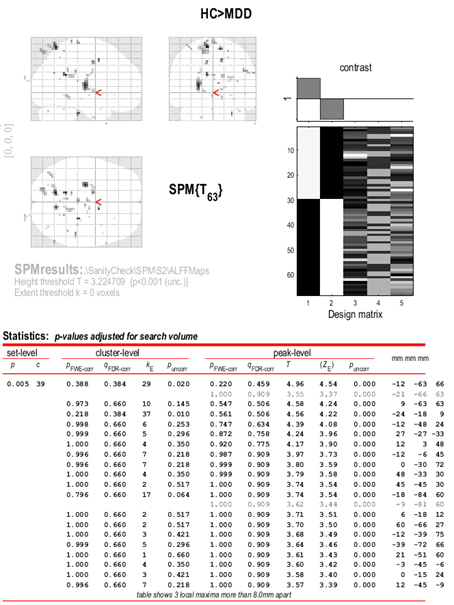
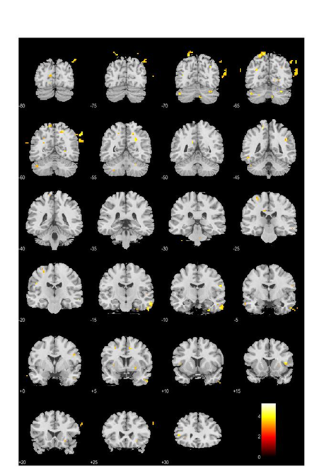
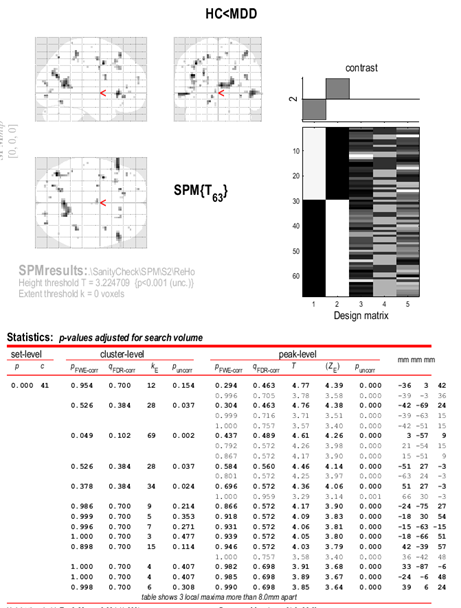
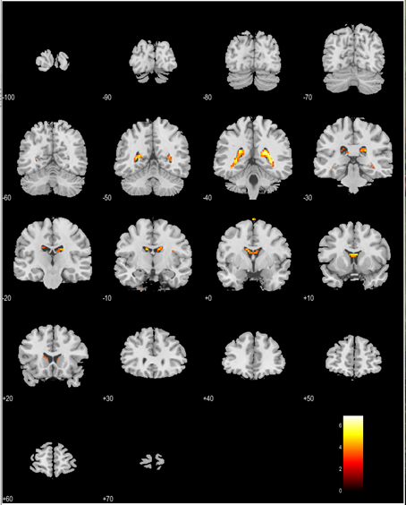
1. I did pre-processing and got the ALFF, fALFF, ReHo results by DPARSF, and then I used zALFF, zfALFF, zReHo files for the 2 sample t-test which I did in the SPM12. But there are some activation regions outside the brain, and the results look very strange. What should I do? Do these results convincing?
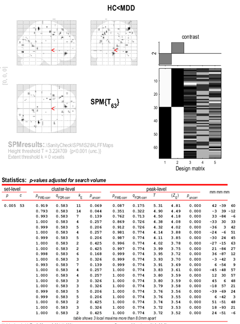
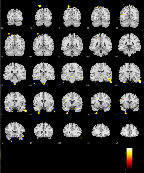
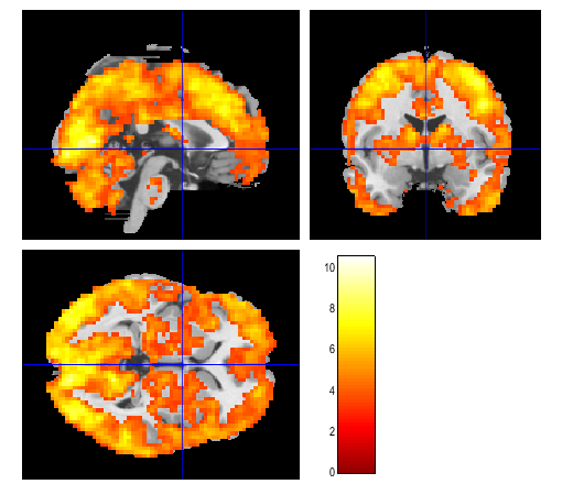
2. I generated two 3*3*3 amygdala masks from AAL altas, because my data is 3*3*3mm, and then I ran the FC analysis in DPARSF. But for the left amygdala FC map two sample t-test (in SPM12 too), I got the activation covered the CSF in one contrast, and the whole brain activation (one cluster) in another contrast. May I ask where do you think the problem could be?
Attached files are the results.
Thanks very much and look forward to your reply.
Best wishes
Yuxi
| Attachment | Size |
|---|---|
| 89.17 KB | |
| 89.03 KB | |
| 226.27 KB | |
| 101.9 KB | |
| 159.54 KB | |
| 87.55 KB | |
| 128.7 KB | |
| 48.26 KB |
Forums:

Powered by Dr. Yan @ My Research Network
YAN Chao-Gan
Mon, 05/18/2020 - 00:54
Permalink
You can use a gray matter
You can use a gray matter mask in statistical analysis.